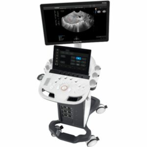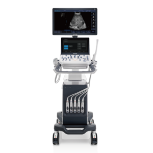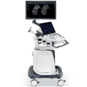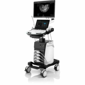


Description
Solution for OB/GYN
P50 has superior image quality, automated measurement tools, and a variety of volume technologies to provide ideal solutions for clinical examinations such as pregnancy examinations, and gynecologic disease diagnosis. With a new 4D transvaginal probe, P50 helps you to see and detect fetal abnormalities, and significantly improves your diagnostic confidence during your examinations.
C-xlasto Imaging
By understanding that tissue stiffness varies depending on the type of tissue, we can use C-xlasto Imaging to easily find abnormalities and tumors within soft tissue. The differences in tissue responses are detected and visualized in real-time by the elastography algorithms through different representations, which can be particularly helpful in analyzing breast, thyroid and musculoskeletal structures.Predominately used only in linear probes, SonoScape’s new transvaginal and bi-plane probe for gynecology and urology are breaking the mold and expanding elastography applications.
Real-time Color Panoramic
With the combination of color flow and real-time panoramic, visualizing the blood flow of an entire vein or artery is now an easy task. Accomplished in real-time for the convenience of the sonographers, any mistakes can also be easily back tracked and corrected without interrupting the scan.
Contrast Imaging
Contrast Imaging on P50 makes full use of the infra harmonic signal and second harmonic signal to improve the image resolution and deep penetration. What’s more, the Dynamic Acoustic Control technology effectively controls the acoustic pressure for the contrast agent, decreasing the required agent dose and assures uniform image quality, guaranteeing longer contrast agent duration and better lesion perfusion of delayed phase observation.
S-Live Silhouette
A unique transparent 3D anatomical image of the fetus for improved initial anatomical review. By using this new application, the system can create completely different fetal images from conventional ultrasound images, which can depict the fetal’s intracorporeal anatomical structure.
Pelvic Floor 4D
Working in conjunction with SonoScape’s latest transvaginal probes, trans-perineal 4D pelvic floor ultrasound provides useful clinical assessment of the impact of vaginal delivery on the female anterior compartment. Allowing doctors to judge whether the pelvic organs prolapsed or not, the extent of porlapse, and determining whether the pelvic muscles tore correctly.
S-Guide
S-Guide gives the user an extensive list of example obstetric ultrasound images as reference guides and a convenient check list system to keep track of their progress during their obstetrics examination.
Auto NT
Auto NT automatically traces and then calculates the thickness of the nuchal posterior transplant layer for a faster evaluation.
Auto Face
Automatically removes masking layers in front of the fetus’s face for a clearer visual of the fetus’s face.
AVC Follicle
AVC Follicle automatically identifies how many follicles are present and calculates their individual volumes.