
Description
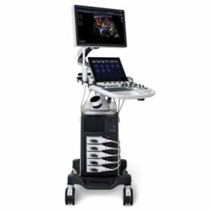
SONOSCAPE E2
Auto Image Optimization
The image is automatically altered and optimized with the click of a button, saving you time with parameter modifications. Furthermore, with Auto Focus enabled, the focus area moves with the depth of the ROI box in the scanning field, giving users with great image quality in the specified area of interest.
Automated Calculation
By automatically tracing the thickness of the carotid vessels, auto IMT is used to determine the level of vascular sclerosis existing in the patient. Auto trace provides customers with sensitive and accurate wave tracing, avoiding manual trace errors and providing calculating results quickly.
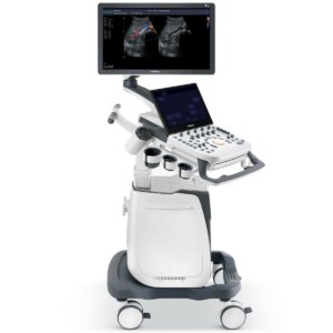
SONOSCAPE E3
Beam Sonoscape E3 C-field
More energy is provided by constantly dynamic focus, which adds to improved contrast resolution, signal-to-noise ratio, and image homogeneity.
μ-Scan
The most recent version of μ-Scan imaging dramatically improves image quality by lowering noise, increasing signal intensity, and improving visibility.
SR Flow
The digital color doppler ultrasound system enables sonographers to easily spot very small veins and slower velocities with SR Flow for the patient’s precise blood flow information.
Tissue Specific Imaging
The device automatically detects different tissues by matching distinct sound ranges, allowing the user to obtain images with greater uniformity and spatial resolution.