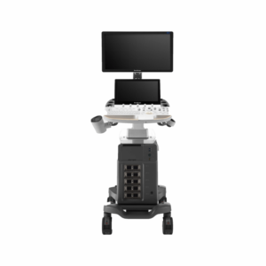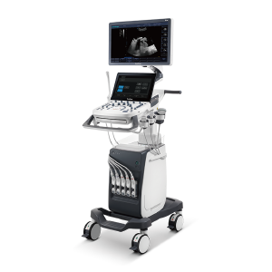


Description
ELITE in General Imaging
μScan+
Available for both B and 3D/4D modes, the new generation μScan+ provides you authentic presentation of details and lesion display through speckle reduction and enhanced border continuity.
SR-Flow
Highly effective filter technology visualizes slow flows, enabling a vivid Doppler display with high sensitivity.
Micro F
Micro F provides an innovative method to expand the range of visible flow in ultrasound, especially for visualizing hemodynamic of tiny vessels.
Bright Flow
3D-like color Doppler flow strengthens boundary definition of vessel walls, without the need of using volume transducer.
CEUS with MFI
Enhanced perfusion display traces small bubble populations, even in low-perfused and peripheral regions.
MFI-Time
To better differentiate tissues, color coded parametric view indicates the uptake time of contrast agents in different perfusion phases.
Strain Elastography
Real-time tissue stiffness assessment based on strain detects potential tissue abnormalities with an intuitive color map displayed. Semi-quantitative analysis of strain ratio indicates relative stiffness of the lesion.
Vis-Needle
Improved accuracy and efficiency in diagnosis are possible with beam steering added to Vis-Needle, which provides enhanced visibility of the needle shaft and needle tip to assist with safe and accurate interventions such as nerve blocks.
ELITE in OB/GYN
S-Live & S-Live Silhouette
S-Live allows for detailed visualization of subtle anatomical features with movable virtual light source, while S-Live Silhouette provides a unique transparent volume image for a more comprehensive internal and external view of the anatomy, both thereby enabling intuitive diagnosis with real-time 3D images and enriching patient communication.
Color 3D
Available on color and power Doppler mode, Color 3D applies advanced rendering, including S-Live, S-Live Silhouette, etc., to blood flow to produce more intuitive and natural hemodynamics of vascular networks, like umbilical cords.
S-Fetus
Realize automatic standard plane acquisition and measurement through AI technologies with just one click. Common fetal biometry results are generated, accurately and efficiently.
Auto OB
A deep learning based method helps to obtain fetal biometry efficiently. Consistent results also reduce user-dependent variability.
Auto NT
It provides semi-automatic and standardized measurements of the nuchal translucency thickness in 2D image and reduces operator dependency on the results.
Auto Face
Auto Face optimizes 3D fetal face display for easier anomalies diagnosis, by removing occlusions and artifacts.
AVC Follicle
A volume-data based automatic follicular calculation includes the number and volume for highly efficient follicle analysis.
Pelvic Floor Imaging
Working in conjunction with specialized transvaginal probes, both 2D and volume imaging are available with thorough evaluation in viewing pelvic anatomy like muscles, bladder, uterus, etc.
ELITE in Cardiovascular
Tissue Doppler Imaging (TDI)
TDI uses myocardial Doppler frequency shifts to present an informative view of myocardial movement indicating velocity and direction.
Stress Echo
A straightforward template for clinicians to conduct stress echo exam. Professional wall motion bulls-eye scoring and reporting provides visualized and intuitive results.
Myocardium Quantitative Analysis (MQA)
Precise quantitative measurement on myocardial mechanics is achieved by MQA based on real-time sensitive wall motion tracking. It provides global and regional assessment including strain, strain rate, displacement, velocity, etc.
LVO
P50 Elite’s enhanced capabilities allow for LV opacification during stress exam to enhance discrimination between myocardial tissue and blood pool, providing better visualization of the endocardia border display especially for difficult patients.
Auto EF
Save more time and efforts compared with manual measurement with Auto EF, which calculates ejection fraction based on left ventricular wall tracing and Simpson’s rule.
Auto IMT
Auto IMT makes the measurement of anterior and posterior intima-media thickness much easier with simple placement of the ROI.
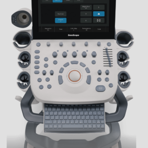
ELITE in Design & User Interaction
Sono-Help
Sono-Help is an inspiring tutorial displaying probe placement, anatomy illustration and standard ultrasound image examples. As a useful reference less experienced clinicians could rely on, Sono-Help covers a variety of applications including liver, kidney, cardiac, breast, thyroid, obstetrics, vascular, etc.
Sono-Assistant
Sono-assistant guides clinicians through the entire exam and provides customizable scanning protocol helps streamline workflow while increasing standardization and reducing keystrokes and exam time.
Sono-Drop
Sono-drop provides a fast and convenient ultrasound image transmission between P50 Elite and the patients’ smart devices. The bond between clinicians and patients are supposed to be strengthened through more frequent communication.
Sono-Synch
Real-time interface and camera sharing, enabled by Sono-synch, makes it possible to connect two ultrasound in a remote distance and perform remote medical consultation and tutorial.